45 compound microscope diagram without labels
Compound Microscope: Definition, Diagram, Parts, Uses, Working ... - BYJUS A compound microscope is defined as A microscope with a high resolution and uses two sets of lenses providing a 2-dimensional image of the sample. The term compound refers to the usage of more than one lens in the microscope. Also, the compound microscope is one of the types of optical microscopes. Label the microscope - Science Learning Hub In this interactive, you can label the different parts of a microscope. Use this with the Microscope parts activity to help students identify and label the main parts of a microscope and then describe their functions. Drag and drop the text labels onto the microscope diagram.
Diagram of a Compound Microscope - Biology Discussion A bright-field or compound microscope is primarily used to enlarge or magnify the image of the object that is being viewed, which can not otherwise be seen by the naked eye. Magnification may be defined as the degree of enlargement of the image of an object provided by the microscope.

Compound microscope diagram without labels
Compound Microscope Parts Made Easy List and Diagram of Compound Microscope Parts: Head - The head is the uppermost part of the microscope that contains the eyepiece, tube, objective lens, and nosepiece. So all the optical parts of a compound microscope are in the head. Eyepiece - The eyepiece is the lens at the top, and the part you look through when using the microscope. Parts of the Microscope with Labeling (also Free Printouts) Parts of the Microscope with Labeling (also Free Printouts) A microscope is one of the invaluable tools in the laboratory setting. It is used to observe things that cannot be seen by the naked eye. Table of Contents 1. Eyepiece 2. Body tube/Head 3. Turret/Nose piece 4. Objective lenses 5. Knobs (fine and coarse) 6. Stage and stage clips 7. Aperture PDF Parts of a Microscope Printables - Homeschool Creations typical student microscope -other microscopes will vary) •Which part of the microscope rotates so another person can look through the eyepiece without needing to move the microscope ? the head •What is the magnification level on the eyepiece of a microscope?10x (see objective lens magnification to see how these work together)
Compound microscope diagram without labels. PDF Label compound microscope worksheet [clearBoth] [clearBoth] Microscope diagram without label After you've studied all the pieces of the composite microscope, it's time to put your brain to the test. Print an unmarked microscope chart and check that you can fill out all the blanks. [clearBoth] [clearBoth] Blank microscope diagram Next we have an empty microscope diagram. Compound Microscope- Definition, Labeled Diagram, Principle, Parts, Uses Alternatively, the magnification of the compound microscope is given by: m = D/ fo * L/fe where, D = Least distance of distinct vision (25 cm) L = Length of the microscope tube fo = Focal length of the objective lens fe = Focal length of the eye-piece lens Parts of a Compound Microscope Eyepiece And Body Tube. Compound Microscope Parts, Functions, and Labeled Diagram Compound Microscope Definitions for Labels. Eyepiece (ocular lens) with or without Pointer: The part that is looked through at the top of the compound microscope. Eyepieces typically have a magnification between 5x & 30x. Monocular or Binocular Head: Structural support that holds & connects the eyepieces to the objective lenses. Compound microscope - their parts and function - Microscopy4kids Compound microscopes have more than one lens to generate high magnification images of flat, thin specimens. 2. Eyepiece (10x) and Objective lenses (4x, 10x, 40x, 100x) are two major optical parts of a microscope. 3. Total magnification power is calculated by multiplying the magnification of the eyepiece and objective lens. 4.
Compound Microscope - Types, Parts, Diagram, Functions and Uses A compound microscope captures an inverted image of the specimen because every time the light passes through the lens, the image's direction is flipped. The image always ends up inverted from the original. So, if you move the sample to the left, it moves in the right direction. Image 18: A comparison image between a simple and compound microscope. What is a Compound Microscope? - New York Microscope Company What is a Compound Microscope? A compound microscope is an instrument that is used to view magnified images of small specimens on a glass slide. It can achieve higher levels of magnification than stereo or other low power microscopes and reduce chromatic aberration. It achieves this through the use of two or more lenses in the objective and the ... Parts of the Compound Microscope - HCC Learning Web Parts of the Compound Microscope Use Figure 2 as a guide to locate the major parts of the compound microscope. a. Base: The bottom, flat part that supports the microscope. b. Arm: The straight or curved vertical part that connects the base to the upper portion. c. Body Tube: Extends from the arm and contains the ocular lens and the rotating 16 Parts of a Compound Microscope: Diagrams and Video Once you have an understanding of the parts of the microscope it will be much easier to navigate around and begin observing your specimen, which is the fun part! The 16 core parts of a compound microscope are: Head (Body) Arm Base Eyepiece Eyepiece tube Objective lenses Revolving Nosepiece (Turret) Rack stop Coarse adjustment knobs
Parts of a Compound Microscope (And their Functions) A compound microscope is the most common microscope you can get and the type you'll typically see in a lab or hobbyist's study. These microscopes tend to have total magnification between 40x - 2000x to allow you to see specimens like bacteria and cells. Compound Microscope Parts - Labeled Diagram and their Functions - Rs ... The term "compound" refers to the microscope having more than one lens. Basically, compound microscopes generate magnified images through an aligned pair of the objective lens and the ocular lens. In contrast, "simple microscopes" have only one convex lens and function more like glass magnifiers. Parts of a Compound Microscope and Their Functions Compound microscope magnification is determined by multiplying the eyepiece and objective powers. When viewed through a 5X eyepiece with a 10X objective, an item is magnified 5 x 10=50 times. The magnification is 10 x 45 = 450 times when using a 10X eyepiece and a 45X objective. How to Use the Compound Microscope Binocular Microscope Anatomy - Parts and Functions with a Labeled Diagram Now, I will discuss the details anatomy of the light compound microscope with the labeled diagram. Why it is called binocular: because it has two ocular lenses or an eyepiece on the head that attaches to the objective lens, this ocular lens magnifies the image produced by the objective lens. Binocular microscope parts and functions
The figure shows a picture of our custom made two-photon microscope | Download Scientific Diagram
A Study of the Microscope and its Functions With a Labeled Diagram Compound Microscope Diagram The compound microscope uses light for illumination. Some compound microscopes make use of natural light, whereas others have an illuminator attached to the base. The specimen is placed on the stage and observed through different lenses of the microscope, which have varying magnification powers.

Labeled Compound Microscope Diagram - Aflam-Neeeak
PDF An Introduction to The Compound Microscope microscope in an upright position using both hands. **When carrying the microscope, place one hand on the base and the other hand around the arm. **DO NOT PLACE THE MICROSCOPE IN AN UPSIDE DOWN POSITION. PIECES WILL FALL OUT. **Keep microscope away from the edge of the bench, particularly when not in use. **Make sure power cords are out of the way.

A Compound Microscope Diagram - Micropedia
Parts of a microscope with functions and labeled diagram Figure: Diagram of parts of a microscope There are three structural parts of the microscope i.e. head, base, and arm. Head - This is also known as the body. It carries the optical parts in the upper part of the microscope. Base - It acts as microscopes support. It also carries microscopic illuminators.
Using the Microscope
Working Principle and Parts of a Compound Microscope (with Diagrams) It holds the stage, body tube, fine adjustment and coarse adjustment. 5. Body Tube: It is usually a vertical tube holding the eyepiece at the top and the revolving nosepiece with the objectives at the bottom. The length of the draw tube is called 'mechanical tube length' and is usually 140-180 mm (mostly 160 mm). 6.

8 Best Images of Lens Diagram Worksheet - Microscope with Labeled Parts, Label Eye Parts ...
Compound Microscope: Parts of Compound Microscope - BYJUS The parts of the compound microscope can be categorized into: Mechanical parts; Optical parts (A) Mechanical Parts of a Compound Microscope. 1. Foot or base. It is a U-shaped structure and supports the entire weight of the compound microscope. 2. Pillar. It is a vertical projection. This stands by resting on the base and supports the stage. 3. Arm

Optical microscope - Wikipedia
PDF Basic Observation Procedures for Compound Microscopes 3. Rotate the 100X objective into position without getting the 40X objective in the oil. 4. While observing from one side of the stage, slowly, raise the stage until you see the meniscus of the oil on the specimen come in contact with the tip of the 100X objective. Now go to the eyepieces and observe as you finish focusing with the fine focus knob.
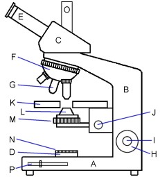
Diagram Of A Microscope With Labels - Drivenhelios
Compound Light Microscope Diagram Worksheet - Google Groups A test over there light microscopes The first 12 questions are on labeling the parts of a microscope and questions 13-20 are multiple. Which microscope diagram worksheet. Students need they turn in source more on microscope when not please use. Get tips on custom to use joint compound microscope see a diagram of the parts of a.

Compound Microscope Parts, Functions, and Labeled Diagram - New York Microscope Company
Microscope Parts and Functions Microscope Parts and Functions With Labeled Diagram and Functions How does a Compound Microscope Work?. Before exploring microscope parts and functions, you should probably understand that the compound light microscope is more complicated than just a microscope with more than one lens.. First, the purpose of a microscope is to magnify a small object or to magnify the fine details of a larger ...
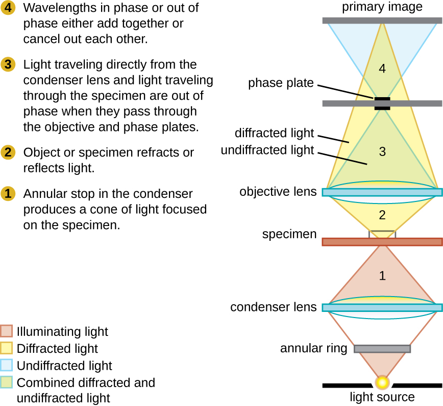
Phase-contrast microscopes By OpenStax (Page 4/16) | QuizOver.com
Labeling the Parts of the Microscope | Microscope World Resources Labeling the Parts of the Microscope This activity has been designed for use in homes and schools. Each microscope layout (both blank and the version with answers) are available as PDF downloads. You can view a more in-depth review of each part of the microscope here. Download the Label the Parts of the Microscope PDF printable version here.

Chapter 1 Questions PPT - BIOLOGY JUNCTION
Labelled Diagram of Compound Microscope - Biology Discussion The below mentioned article provides a labelled diagram of compound microscope. Part # 1. The Stand: The stand is made up of a heavy foot which carries a curved inclinable limb or arm bearing the body tube. The foot is generally horse shoe-shaped structure (Fig. 2) which rests on table top or any other surface on which the microscope in kept.

The Compound Microscope Worksheets | Microscope, Biology lessons, Anatomy and physiology book
Microscope, Microscope Parts, Labeled Diagram, and Functions Revolving Nosepiece or Turret: Turret is the part of the microscope that holds two or multiple objective lenses and helps to rotate objective lenses and also helps to easily change power. Objective Lenses: Three are 3 or 4 objective lenses on a microscope. The objective lenses almost always consist of 4x, 10x, 40x and 100x powers. The most common eyepiece lens is 10x and when it coupled with ...
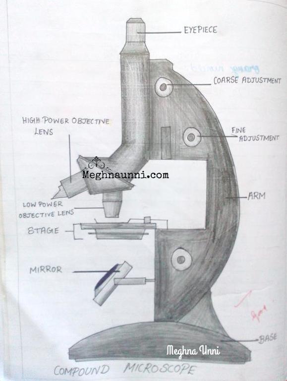
Meghna Unni's Blog: Meghna's Paintings, Bharathanatyam, Writings, Stories, Projects, Crafts
PDF Parts of a Microscope Printables - Homeschool Creations typical student microscope -other microscopes will vary) •Which part of the microscope rotates so another person can look through the eyepiece without needing to move the microscope ? the head •What is the magnification level on the eyepiece of a microscope?10x (see objective lens magnification to see how these work together)

Microscope World Blog: High School Microscope Features
Parts of the Microscope with Labeling (also Free Printouts) Parts of the Microscope with Labeling (also Free Printouts) A microscope is one of the invaluable tools in the laboratory setting. It is used to observe things that cannot be seen by the naked eye. Table of Contents 1. Eyepiece 2. Body tube/Head 3. Turret/Nose piece 4. Objective lenses 5. Knobs (fine and coarse) 6. Stage and stage clips 7. Aperture
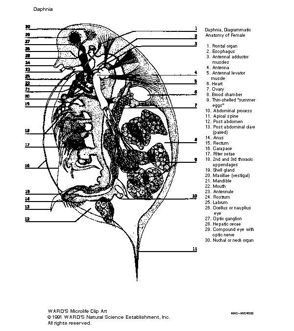
Daphnia Labeled Diagram
Compound Microscope Parts Made Easy List and Diagram of Compound Microscope Parts: Head - The head is the uppermost part of the microscope that contains the eyepiece, tube, objective lens, and nosepiece. So all the optical parts of a compound microscope are in the head. Eyepiece - The eyepiece is the lens at the top, and the part you look through when using the microscope.

Microscope Clip Art at Clker.com - vector clip art online, royalty free & public domain
Post a Comment for "45 compound microscope diagram without labels"