39 eye diagram and labels
What Does the Eye Look Like? - Diagram of the Eye | Harvard Eye Associates Vitreous Gel: A thick, transparent liquid that fills the center of the eye. It is mostly water and gives the eye its form and shape. Our eyes are vital for seeing the world around us. Keep them healthy by maintaining regular vision exams. Contact Harvard Eye Associates at 949-951-2020 or harvardeye.com to schedule an appointment today. eye labeling Diagram | Quizlet sclera. Tough white out covering of the eyeball. choroid. Middle layer of the eye (between the retina and the sclera) that contains the blood vessels that nourish the eye and cornea. iris. colored layer that dilates and constricts to allow in more or less light. ciliary body. structure on each side of the lens that connects the choroid and iris.
Human Eye Ball Anatomy & Physiology Diagram - eMedicineHealth The orbit is the bony eye socket of the skull. The orbit is formed by the cheekbone, the forehead, the temple, and the side of the nose. The eye is cushioned within the orbit by pads of fat. In addition to the eyeball itself, the orbit contains the muscles that move the eye, blood vessels, and nerves. The orbit also contains the lacrimal gland ...
Eye diagram and labels
Anatomy of the eye: Quizzes and diagrams | Kenhub One of our favorite ways to get to grips with all of the parts of the eye is by utilizing labeled diagrams. On a diagram of the eye, we can see all of the relevant structures together on one image. This helps us to understand how each one is situated and related to the other. Labeled diagram of the eye Labeled Eye Diagram | Eye anatomy diagram, Eye anatomy, Diagram of the eye Labeled Eye Diagram. Labeled Eye Diagram. Samer kursa. 16 followers. Eye Anatomy Diagram. Human Eye Diagram. Diagram Of The Eye. Brain Anatomy. Anatomy And Physiology. Human Anatomy ... labeled eye model. Jean Ruddell. I BIO 1147/1121 Laboratory. Similar ideas popular now. Photography. Subjects. Human eye - Wikipedia Each eye has seven extraocular muscles located in its orbit. Six of these muscles control the eye movements, the seventh controls the movement of the upper eyelid.The six muscles are four recti muscles – the lateral rectus, the medial rectus, the inferior rectus, and the superior rectus, and two oblique muscles the inferior oblique, and the superior oblique.
Eye diagram and labels. Labelling the eye — Science Learning Hub In this interactive, you can label parts of the human eye. Use your mouse or finger to hover over a box to highlight the part to be named. Drag and drop the text labels onto the boxes next to the eye diagram If you want to redo an answer, click on the box and the answer will go back to the top so you can move it to another box. North Carolina Eye Care & Eye Exams | eyecarecenter North Carolina Eye Care Services. The eye doctors at eyecarecenter are here to help you with all of your eye care needs. Our highly trained eye care professionals focus on maintaining your eye health with comprehensive eye care, preventative care, and treatment. Trust us for routine checkups and treating conditions like glaucoma, cataracts & more. Eye pattern - Wikipedia In telecommunication, an eye pattern, also known as an eye diagram, is an oscilloscope display in which a digital signal from a receiver is repetitively sampled and applied to the vertical input, while the data rate is used to trigger the horizontal sweep. It is so called because, for several types of coding, the pattern looks like a series of eyes between a pair of rails. diagram of eye with labels Horseshoe Crab Anatomy. 16 Pics about Horseshoe Crab Anatomy : Label the Eye, Eye With Labels Clip Art at Clker.com - vector clip art online, royalty and also Muscles of the Human Eyeball | ClipArt ETC. Horseshoe Crab Anatomy dnr.maryland.gov crab horseshoe anatomy eyes diagram labeled gills ccs dnr maryland gov
Male Human Anatomy Diagram Pictures, Images and Stock Photos Labeled Anatomy Chart of Male Muscles on White Background Labeled human anatomy diagram of man's full body muscular system from a posterior view on a white background. male human anatomy diagram stock pictures, royalty-free photos & images The Human Eye (Eyeball) Diagram, Parts and Pictures The wall of the eyeball is made up of three layers - fibrous (outer), vascular/muscular (middle) and sensorineural (inner) layers. Diagram of the different layers of the eyeball Outer Layer The outer fibrous layer maintains the shape of the eyeball and protects more fragile internal structure. This layer is made up of the sclera and cornea. Eye Anatomy: A Closer Look At the Parts of the Eye - All About Vision Eye anatomy: A closer look at the parts of the eye. When surveyed about the five senses — sight, hearing, taste, smell and touch — people consistently report that their eyesight is the mode of perception they value (and fear losing) most. Despite this, many people don't have a good understanding of the anatomy of the eye, how vision works ... Eye Diagram With Labels and detailed description - BYJUS A brief description of the eye along with a well-labelled diagram is given below for reference. Well-Labelled Diagram of Eye The anterior chamber of the eye is the space between the cornea and the iris and is filled with a lubricating fluid, aqueous humour. The vascular layer of the eye, known as the choroid contains the connective tissue.
Diagram of the Eye - Lions Eye Institute Instructions Click the parts of the eye to see a description for each. Hover the diagram to zoom. Need any help? If you would like to know more about us, or want to make an appointment, please don't hesitate to get in touch. (08) 9381 0777 carecentre@lei.org.au Request an appointment Customer Care Centre (08) 9381 0777 Human eye diagram to label - simplediagram.netlify.app Human eye diagram labeled parts of the human eye diagram and human eye diagram labeled are three main things we want to present to you based on the post title. Layer of cells on the back of the eye. Controls how much light enters the eye. To play the game online visit Labeling Parts of the Eye 5th Grade. In the 2nd worksheet they match the. PDF Parts of the Eye - National Institutes of Health Eye Diagram Handout Author: National Eye Health Education Program of the National Eye Institute, National Institutes of Health Subject: Handout illustrating parts of the eye Keywords: parts of the eye, eye diagram, vitreous gel, iris, cornea, pupil, lens, optic nerve, macula, retina Created Date: 12/16/2011 12:39:09 PM Human Eye Diagram, How The Eye Work -15 Amazing Facts of Eye The shark has even been used in human eye surgery! FACT 4 The length of our eyes are about 1 inch across and weigh about 0.25 ounce. FACT 5 Our eyeballs stay the same size forever but our nose and ears continue to grow. FACT 6 Eyes are the second most complex organ after the brain.
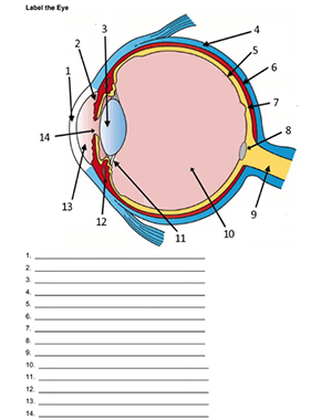
Label the Eye
Labelled Diagram of Human Eye, Explanation and Function - VEDANTU Labeled Diagram of Human Eye The eyes of all mammals consist of a non-image-forming photosensitive ganglion within the retina which receives light, adjusts the dimensions of the pupil, regulates the availability of melatonin hormones, and also entertains the body clock.

Human eye anatomy stock vector. Illustration of diagram - 37354087
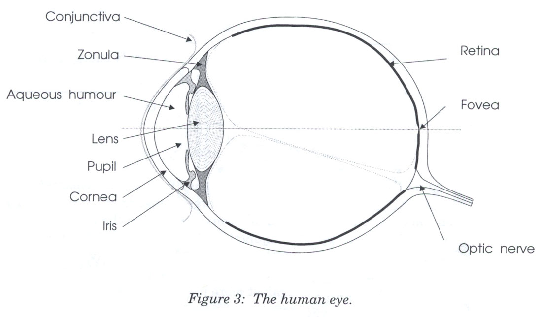
Eye Anatomy: 16 Parts of the Eye & Their Functions - Vision Center The following are parts of the human eyes and their functions: 1. Conjunctiva The conjunctiva is the membrane covering the sclera (white portion of your eye). The conjunctiva also covers the interior of your eyelids. Conjunctivitis, often known as pink eye, occurs when this thin membrane becomes inflamed or swollen.
31 Label The Parts Of Eye - Labels 2021
Generate eye diagram - MATLAB eyediagram - MathWorks eyediagram(x,n) generates an eye diagram for signal x, plotting n samples in each trace. The labels on the horizontal axis of the diagram range between –1/2 and 1/2. The function assumes that the first value of the signal and every nth value thereafter, occur at integer times.
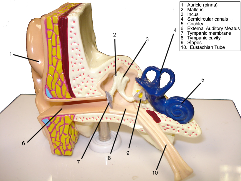
Eye and Ear Models
Structure and Functions of Human Eye with labelled Diagram - BYJUS Structure and Functions of Human Eye with labelled Diagram Biology Biology Article Structure Of Eye Structure of the Eye The eye is one of the sensory organs of the body. In this article, we shall explore the anatomy of the eye The structure of the eye is an important topic to understand as it one of the important sensory organs in the human body.
Diagram Human Eye Labeled - Aflam-Neeeak
Diagram Maker | Online Diagramming and Design Solution - Venngage Create eye-catching, informative diagrams without any design experience. Choose from a range of diagram templates to get started. Each diagram template is endlessly customizable, so you can make it as complex, concise or creative as you like. Venngage's free diagram maker lets you create engaging diagrams using unique icons and illustrations.

Perception: 3.1 Eye to brain - YouTube
Eye Anatomy: Parts of the Eye and How We See Eye Anatomy: Parts of the Eye Outside the Eyeball The eye sits in a protective bony socket called the orbit. Six extraocular muscles in the orbit are attached to the eye. These muscles move the eye up and down, side to side, and rotate the eye. The extraocular muscles are attached to the white part of the eye called the sclera.
Reading 15: Color
Eye labeling Diagram | Quizlet Female reproductive system labeled. 26 terms. danni_dirato. 12 steps of blood flow. 12 terms. Nicky-De. Sets with similar terms. Eye Anatomy. 14 terms. bcnelson211 TEACHER. Anatomy - Eye Vocabulary. 16 terms. vickyttoriaa. AP Psychology Eye diagram. 8 terms. jack_rowen. Sheep Eye Dissection. 19 terms. JENNIFER_WOOD9. Other sets by this creator.

Pin on Examples Printable Label Templates
PDF Eye Anatomy Handout - National Institutes of Health Eye Anatomy Handout Author: National Eye Institute , National Eye Health Education Program Subject: Diabetes and Healthy Eyes Toolkit and Website Keywords: Eye anatomy, eye diagram, cornea, iris, lens, macula, optic nerve, pupil, retina, vitrous gel, diabetic eye disease. Created Date: 6/27/2012 11:57:40 AM
Unlabelled Respiratory System Clip Art at Clker.com - vector clip art online, royalty free ...
Eye diagram basics: Reading and applying eye diagrams - EDN Eye diagrams usually include voltage and time samples of the data acquired at some sample rate below the data rate. In Figure 1, the bit sequences 011, 001, 100, and 110 are superimposed over one another to obtain the final eye diagram. Figure 1These diagrams illustrate how an eye diagram is formed.
Eye Anatomy Diagramvector Illustration Stock Vector (Royalty Free) 485878195 - Shutterstock
Label Parts of the Human Eye - University of Dayton Parts of the Eye. Select the correct label for each part of the eye. The image is taken from above the left eye. Click on the Score button to see how you did. Incorrect answers will be marked in red. ...

Eye Diagram Without Labels | via Anatomy Pictures Gallery if… | Flickr
Eye diagram labeled - Healthiack Eye diagram labeled This summary post is displaying Eye diagram labeled … The eyes are responsible for our sense of sight or vision. Any disruption of the anatomy and physiology of the eyes and their supporting structures can cause vision impairment. Some of the ocular disorders or medical conditions affecting the eyes include the following:
Neuron B&w Clip Art at Clker.com - vector clip art online, royalty free & public domain
6,819 Human eye diagram Images, Stock Photos & Vectors - Shutterstock Find Human eye diagram stock images in HD and millions of other royalty-free stock photos, illustrations and vectors in the Shutterstock collection. Thousands of new, high-quality pictures added every day.
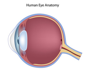
Diagram Of The Eye Not Labeled - Diagram Media
Free Venn Diagram Maker by Canva Venn diagram maker features. Canva’s Venn diagram maker is the easiest way to make a Venn diagram online. Start by choosing a template – we’ve got hundreds of Venn diagram examples to choose from. With a suite of easy to use design tools, you have complete control over the way it looks.

Picture Of the Eye Labeled Elegant Human Eye Anatomy for Kids | Human eye diagram, Eye anatomy ...
Labeled Eye Diagram | Science Trends What you want to interpret as a major part of the human eye is somewhat up to the individual, but in general there are seven parts of the human eye: the cornea, the pupil, the iris, the lens, the vitreous humor, the retina, and the sclera. Let's take a closer look at each of these components individually. The Cornea
Label the parts of the eye: | bartleby
Eye Diagram Teaching Resources | Teachers Pay Teachers The Human Eye Overview Reading Comprehension and Diagram Worksheet. by. Teaching to the Middle. 4.7. (65) $1.50. Zip. This passage briefly describes the human eye (900-1000 Lexile). 14 questions (matching and multiple choice) assess students' understanding. Students label a diagram of 6 parts of the eye.
Post a Comment for "39 eye diagram and labels"