41 eye diagram with labels and functions
Cow's Eye Dissection - Eye diagram - Exploratorium The pupil is the dark circle in the center of your iris. It's a hole that lets light into the inner eye. Your pupil is round. A cow's pupil is oval. A tough, clear covering over the iris and the pupil that helps protect the eye. Light bends as it passes through the cornea. This is the first step in making an image on the retina. diagram of eye with labelling Eye Diagram With Labels And Functions - Aflam-Neeeak aflam-neeeak.blogspot.com Earthworm Presentation earthworm labeled dissection external anatomy labels presentation lumbricus label section cross slideshare sp fig physiology Labeled Eye Diagram ks2 links markcritz Human Eye Anatomy Quiz
Labelling the eye — Science Learning Hub The human eye contains structures that allow it to perceive light, movement and colour differences. In this activity, students use online or paper resources to identity and label the main parts of the human eye. By the end of this activity, students should be able to: identify the main parts of the human eye

Eye diagram with labels and functions
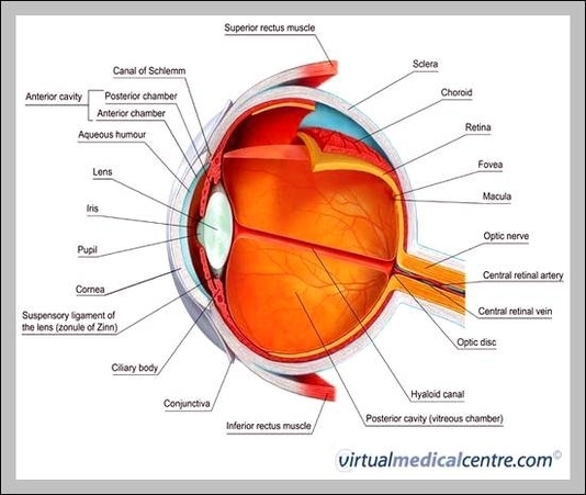
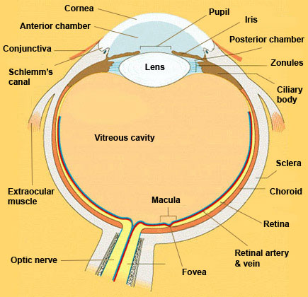
Label Parts of the Human Eye - University of Dayton Parts of the Eye. Select the correct label for each part of the eye. The image is taken from above the left eye. Click on the Score button to see how you did. Incorrect answers will be marked in red. ... Human Eye Ball Anatomy & Physiology Diagram - eMedicineHealth The orbit is the bony eye socket of the skull. The orbit is formed by the cheekbone, the forehead, the temple, and the side of the nose. The eye is cushioned within the orbit by pads of fat. In addition to the eyeball itself, the orbit contains the muscles that move the eye, blood vessels, and nerves. The orbit also contains the lacrimal gland ... Structure of Human Eye (With Diagram) | Human Body ADVERTISEMENTS: In this article we will discuss about the structure of human eye. Structure of Human Eye: The eye is a hollow, spherical structure measuring about 2.5 cm in diameter. ADVERTISEMENTS: Its wall is composed of three coats: 1. The outer fibrous coat— sclera, cornea. 2. The middle vascular coat— choroid, ciliary body, iris. 3. […]
Eye diagram with labels and functions. Structure and Functions of Human Eye with labelled Diagram Structure and Functions of Human Eye with labelled Diagram Biology Biology Article Structure Of Eye Structure of the Eye The eye is one of the sensory organs of the body. In this article, we shall explore the anatomy of the eye The structure of the eye is an important topic to understand as it one of the important sensory organs in the human body. Labelled Diagram of Human Eye, Explanation and Function - VEDANTU The basic functions of Rods and Cones are conscious light perception, color differentiation and depth perception. The human eye is capable of distinguishing between about 10 million colors, and it can also detect a single photo. The human eye is a part of the sensory nervous system. Labeled Diagram of Human Eye Human Ear Diagram Without Labels - picture front of the eye without ... Human Ear Diagram Without Labels - 18 images - datei anatomy of the human ear svg wikipedia, ear anatomy diagram, circulatory system diagram without labels fresh smorgasbord variety, unlabeled digestive system diagram without labels news word, Labelling the eye — Science Learning Hub In this interactive, you can label parts of the human eye. Use your mouse or finger to hover over a box to highlight the part to be named. Drag and drop the text labels onto the boxes next to the eye diagram If you want to redo an answer, click on the box and the answer will go back to the top so you can move it to another box.
MCAT Eye Anatomy: Eye Structure & Function - Magoosh MCAT Blog MCAT Eye Anatomy: Diagram of the Human Eye Light refracts (bends) as it passes sequentially through the cornea, aqueous humor, lens, and vitreous humor. Errors in refraction cause visual defects which can be corrected by contacts or glasses. Myopia and hyperopia are two types of refractive error. The Human Eye - Diagram, Parts, Working, Function and Work of ... - VEDANTU The human eye operates similar to a digital camera in several ways: Light focuses mainly on the cornea, which acts like a camera lens. The iris controls the light that reaches the eye by adjusting the size of the pupil, and thus it functions like the diaphragm of a camera. The lens of the eye is located behind the pupil, and it focuses light. Labeled Eye Diagram - Pinterest This vibrant 20" x 26" (51 x 66 cm) exam-room anatomy poster shows cross section of The Eye. It also provides lateral and superior view of the eye and shows the visual field. Anterior chamber angle, eyelashes, tear ducts, cornea, lens, retina, fundus and the macula lutea are illustrated. Human Eye Diagram, How The Eye Work -15 Amazing Facts of Eye First, light rays enter the eye through the cornea, the clear front "window" of the eye. The dome shaped cornea bends light to help the eye focus. From the cornea, the light passes through an opening called the pupil. The amount of light passing through is controlled by the iris, or the colored part of your eye.
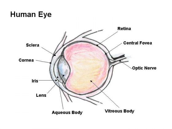
Diagram of the Eye - Home - Lions Eye Institute To understand the eye and its functions, it's important to understand how the eye works, see below diagrams for both the external eye and the internal eye. The External Eye Instructions Click the parts of the eye to see a description for each. Hover the diagram to zoom. The Internal Eye Instructions Eye Anatomy | Definition, Structure & Functions - iBiologia Diagram of Human Eye with Labelling. Eye Anatomy Complete Physiology of Eye is described below in the given paragraph: The eye is rather like a living Camera. Each eye is a liquid-filled ball 2.5 cm in diameter. At the front of the eye is a clear, round window called the cornea. Behind the cornea is a "lens. Eye Anatomy: 16 Parts of the Eye & Their Functions The following are parts of the human eyes and their functions: 1. Conjunctiva The conjunctiva is the membrane covering the sclera (white portion of your eye). The conjunctiva also covers the interior of your eyelids. Conjunctivitis, often known as pink eye, occurs when this thin membrane becomes inflamed or swollen. Eye anatomy and function - AboutKidsHealth For people with normally functioning eyes, the following sequence takes place: Light reflects off the object we are looking at. Light rays enter the eye through the cornea at the front of the eye. The light passes through a watery fluid (aqueous humor), and enters the pupil to reach the lens.
Diagram Of Inside Of Mouth - Mature Milf
PDF Eye Anatomy Handout - National Institutes of Health of light entering the eye. Lens: The lens is a clear part of the eye behind the iris that helps to focus light, or an image, on the retina. Macula: The macula is the small, sensitive area of the retina that gives central vision. It is located in the center of the retina. Optic nerve: The optic nerve is the largest sensory nerve of the eye.

SOLVED:Label this diagram of a human eye.
Labeled Eye Diagram | Human eye diagram, Eye anatomy ... - Pinterest Science Notes. CONTENTSEyesVideo: Anatomy and Function of the EyeEarsVideo: Ear Anatomy Our most important sensory receptors are the eyes and the ears. The eye is the primary organ for sight, and the ear is the primary organ for sound and equilibrium. Obviously, any impairment of either of these sensory receptors can be a traumatic experience ...

Eye diagram labeled
The Eyes (Human Anatomy): Diagram, Optic Nerve, Iris, Cornea ... - WebMD The front part (what you see in the mirror) includes: Iris: the colored part. Cornea: a clear dome over the iris. Pupil: the black circular opening in the iris that lets light in. Sclera: the ...
Anatomy – Page 32 – Graph Diagram
Eye Anatomy: Parts of the Eye and How We See Behind the anterior chamber is the eye's iris (the colored part of the eye) and the dark hole in the middle called the pupil. Muscles in the iris dilate (widen) or constrict (narrow) the pupil to control the amount of light reaching the back of the eye. Directly behind the pupil sits the lens. The lens focuses light toward the back of the eye.
Eye diagram labeled
Eye anatomy: A closer look at the parts of the eye The iris of the eye functions like the diaphragm of a camera, controlling the amount of light reaching the back of the eye by automatically adjusting the size of the pupil (aperture). The eye's crystalline lens is located directly behind the pupil and further focuses light.

LABELLED EYE DIAGRAM - YouTube
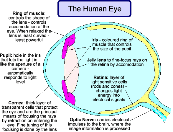
Parts of the Eye and Their Functions - Robertson Opt The iris is the area of the eye that contains the pigment which gives the eye its color. This area surrounds the pupil, and uses the dilator pupillae muscles to widen or close the pupil. This allows the eye to take in more or less light depending on how bright it is around you. If it is too bright, the iris will shrink the pupil so that they ...

Labeled Eye Diagram Gcse - Juanribon.com - ClipArt Best - ClipArt Best
PDF Parts of the Eye - National Eye Institute | National Eye Institute To understand eye problems, it helps to know the different parts that make up the eye and the functions of these parts. Here are descriptions of some of the main parts of the eye: ... Handout illustrating parts of the eye Keywords: parts of the eye, eye diagram, vitreous gel, iris, cornea, pupil, lens, optic nerve, macula, retina ...
health Archives - Page 3 of 3 - Medical Information Illustrated
Eye Anatomy Diagram - EnchantedLearning.com Retina - light-sensitive tissue that lines the back of the eye. It contains millions of photoreceptors (rods and cones) that convert light rays into electrical impulses that are relayed to the brain via the optic nerve. Rods - cells the in the retina that sense brightness (they are photoreceptors). Night vision involves mostly rods (not cones).
Cyberphysics - The human eye, sight defects and their correction
Eye Diagram With Labels and detailed description - BYJUS A brief description of the eye along with a well-labelled diagram is given below for reference. Well-Labelled Diagram of Eye The anterior chamber of the eye is the space between the cornea and the iris and is filled with a lubricating fluid, aqueous humour. The vascular layer of the eye, known as the choroid contains the connective tissue.

Microscope Diagram to Print
Eye Diagram - an overview | ScienceDirect Topics An eye diagram provides a simple and useful tool to visualize intersymbol interference between data bits. Figure 24a shows a perfect eye diagram. A square bit stream (i.e., series of symbol '1's and '0's) is sliced into sub-bit stream with predetermined eye intervals (i.e., several bit periods), and displayed through bit analyzing equipment (e.g., digital channel analyzer), overlapping ...
26 Drag The Labels Onto The Diagram To Identify The Types Of Epithelia. - Wiring Database 2020
The Eye Diagram: What is it and why is it used? The eye diagram is used primarily to look at digital signals for the purpose of recognizing the effects of distortion and finding its source. To demonstrate using a Tektronix MDO3104 oscilloscope, we connect the AFG output on the back panel to an analog input channel on the front panel and press AFG so a sine wave displays. Then we press Acquire.

anatomy, eye diagram to label | Eye anatomy diagram, Eye anatomy, Anatomy
Structure of Human Eye (With Diagram) | Human Body ADVERTISEMENTS: In this article we will discuss about the structure of human eye. Structure of Human Eye: The eye is a hollow, spherical structure measuring about 2.5 cm in diameter. ADVERTISEMENTS: Its wall is composed of three coats: 1. The outer fibrous coat— sclera, cornea. 2. The middle vascular coat— choroid, ciliary body, iris. 3. […]
Post a Comment for "41 eye diagram with labels and functions"